
Dementia diagnosis can be a complex and challenging process for both patients and healthcare professionals. A critical component in this journey is understanding how imaging techniques, particularly CT scans, contribute to accurate identification and treatment planning. When it comes to dementia diagnosis, advanced imaging technologies like CT scans play a crucial role in identifying abnormalities in brain structure and function. By analyzing the findings from these scans, doctors can gain valuable insights into the progression of the disease and develop effective care strategies. This article will explore how CT scan findings aid in diagnosing dementia, including common imaging results and the latest advancements in imaging techniques that enhance diagnostic accuracy. By the end of this article, you’ll have a better understanding of how to interpret CT scan results for an accurate diagnosis and treatment plan.
What is Dementia?
Dementia is a complex and multifaceted condition that affects millions worldwide, and understanding its core characteristics is essential for grasping its relationship with CT scan findings. Let’s start by exploring what dementia actually is.
Definition and Types of Dementia
The most common type of dementia is Alzheimer’s disease, accounting for 60-80% of all cases. It’s characterized by a progressive decline in memory and cognitive abilities, such as problem-solving and judgment. Vascular dementia, on the other hand, occurs when reduced blood flow to the brain causes damage. This can be due to strokes or small vessel disease.
Lewy body dementia is marked by the presence of abnormal protein clumps called Lewy bodies in the brain. These disrupt normal brain function, leading to symptoms like visual hallucinations and difficulty with movement. Frontotemporal dementia affects the front and temporal lobes of the brain, causing changes in personality, behavior, and language skills.
It’s essential to note that each type of dementia has distinct characteristics, but they often share similar cognitive decline. A precise diagnosis typically requires a comprehensive evaluation, including medical history, physical examination, laboratory tests, and imaging studies like CT scans or MRI. While CT scan findings can provide valuable information, they should be interpreted in conjunction with clinical symptoms and other diagnostic tools. Early detection is crucial for effective management and treatment of dementia.
Risk Factors and Causes of Dementia
Age is a significant risk factor for dementia, with most cases occurring after the age of 65. Genetics also play a crucial role, as certain genetic mutations can increase an individual’s likelihood of developing dementia. For example, individuals with a family history of Alzheimer’s disease, particularly those with a first-degree relative affected, are at higher risk.
Underlying medical conditions like hypertension and diabetes can also contribute to the development of dementia. Research suggests that managing these conditions through lifestyle changes and medication can help mitigate this risk. Additionally, certain lifestyle choices, such as physical inactivity, smoking, and excessive alcohol consumption, have been linked to an increased risk of dementia.
Certain medical conditions, including stroke and traumatic brain injury, can also increase the likelihood of developing dementia. In some cases, these conditions may even accelerate the progression of dementia symptoms. For instance, a study found that individuals who experienced a stroke were more likely to develop vascular dementia than those without a history of stroke.
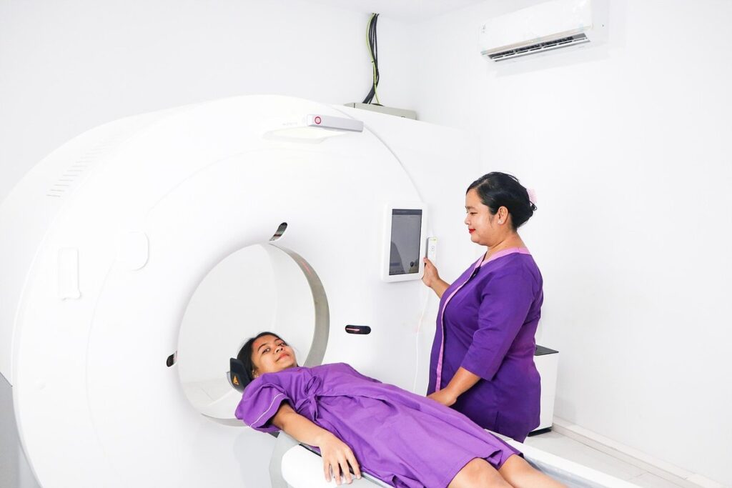
CT Scan Basics: What to Expect
Before undergoing a CT scan, it’s essential to understand what you can expect from the procedure and how it will help doctors assess dementia-related changes in your brain. We’ll break down the basics of CT scans in simple terms.
How a CT Scan Works
To prepare for a CT scan of the brain, you’ll typically be asked to lie on an examination table and slide into the scanner’s opening. The machine is large and circular, with a doughnut-shaped hole in the center where the X-rays will pass through your body. You may be given a contrast agent, usually an iodine-based solution, to help highlight certain areas of the brain.
During the procedure, you’ll remain still while the machine rotates around you. The rotating X-ray beam creates multiple cross-sectional images as it scans the brain from different angles. These images are then reconstructed by computer software into detailed pictures of your brain’s internal structures. The entire process typically takes 2-5 minutes to complete.
The scanner itself is equipped with safety features, such as a backup system and emergency shutdown procedure, in case of any technical issues during the scan. You may also be able to communicate with the technologist through an intercom system if you experience any discomfort or anxiety during the procedure.
Types of CT Scans Used for Dementia Diagnosis
Non-contrast CT scans are commonly used as a first-line imaging modality for dementia diagnosis. This type of scan uses X-rays to produce detailed cross-sectional images of the brain without the use of contrast agents. Non-contrast CT scans can help identify vascular changes, such as white matter hyperintensities, which are often seen in patients with dementia.
Contrast-enhanced CT scans may be ordered to further evaluate suspected conditions like normal pressure hydrocephalus or tumors that could be contributing to cognitive decline. In these cases, a contrast agent is administered intravenously before the scan to help highlight specific areas of interest within the brain.
CT angiography can also be used to assess blood vessel integrity and detect potential vascular causes of dementia, such as stenosis or occlusion. This type of scan uses a combination of X-rays and a rapid bolus injection of contrast agent to create detailed images of cerebral vasculature. The choice of CT scan type depends on the individual patient’s needs and clinical presentation.
Common CT Scan Findings in Dementia Patients
When reviewing a CT scan, healthcare professionals often look for specific patterns and abnormalities that can indicate dementia. We’ll examine some common findings associated with this condition.
Vascular Changes on CT Scans
On a CT scan, vascular changes in dementia patients often appear as white matter hyperintensities and cerebral microbleeds. These lesions are visible because they have different signal intensities than surrounding brain tissue due to their altered composition. White matter hyperintensities, for example, can be caused by demyelination or gliosis, which alter the normal density of these areas. Cerebral microbleeds, on the other hand, indicate small hemorrhages that are often associated with amyloid angiopathy.
The presence of white matter hyperintensities and cerebral microbleeds on a CT scan can indicate vascular dementia or contribute to the development of Alzheimer’s disease. The extent and location of these lesions can provide valuable information about the underlying pathology and help guide further diagnosis. However, it’s essential to note that CT scans may not always detect smaller lesions or those with low density.
To better understand the implications of these findings, consider that a study found that patients with more extensive white matter hyperintensities were at higher risk for cognitive decline. As such, the presence and extent of vascular changes on a CT scan should be carefully evaluated in conjunction with clinical presentation and other diagnostic tests.
Atrophy and Shrinkage of Brain Tissue
On a CT scan, brain atrophy and shrinkage appear as reduced volume of brain tissue. This can manifest as decreased density or contrast between the gray matter and white matter. In patients with dementia, atrophy is often most pronounced in regions critical for memory and cognitive processing, such as the hippocampus and temporal lobe.
The degree of atrophy correlates with the severity of cognitive decline. Studies have shown that patients with more significant atrophy tend to exhibit greater impairments in memory, language, and executive function. Conversely, those with relatively preserved brain volume often experience milder symptoms.
CT scans can also reveal shrinkage of specific brain regions, such as the medial temporal lobe or frontal cortex. This is particularly common in Alzheimer’s disease, where neurodegenerative changes lead to progressive loss of neurons and supporting cells. Radiologists may use established grading scales, like the Scheltens scale, to quantify atrophy severity and correlate it with clinical findings.
Keep in mind that CT scans are not always sensitive enough to detect early-stage atrophy. In these cases, advanced imaging modalities or combination with other diagnostic tools may be necessary for accurate assessment.
Advanced Imaging Techniques for Dementia Diagnosis
Advanced imaging techniques, such as diffusion tensor imaging and magnetic resonance spectroscopy, can help identify specific brain changes associated with dementia. These methods offer valuable insights into disease progression and potential treatment targets.
Functional MRI (fMRI) and Magnetoencephalography (MEG)
Functional MRI (fMRI) measures changes in blood flow to different areas of the brain during various tasks, while magnetoencephalography (MEG) detects magnetic fields generated by electrical activity. These techniques allow researchers and clinicians to study brain function in greater detail than possible with structural imaging alone.
In dementia diagnosis, fMRI is often used to assess cognitive functions such as memory, language, and problem-solving. For example, studies have shown that people with Alzheimer’s disease exhibit altered patterns of brain activity during memory recall tasks. MEG, on the other hand, can provide high-resolution maps of neural activity, which can help identify specific brain regions involved in dementia-related symptoms.
While fMRI and MEG are primarily research tools, they may also be used in clinical settings to support diagnosis or monitor disease progression. However, these techniques require specialized equipment and expertise, limiting their accessibility for routine diagnostic purposes. Despite this limitation, integrating fMRI and MEG findings into a comprehensive diagnostic workup can provide valuable insights into the underlying mechanisms of dementia-related brain dysfunction.
Diffusion Tensor Imaging (DTI) and Tract-Based Spatial Statistics (TBSS)
Diffusion Tensor Imaging (DTI) is a magnetic resonance imaging (MRI) technique that measures water diffusion in the brain’s white matter tracts. In patients with dementia, DTI can detect subtle alterations in white matter integrity and tract structure. By analyzing the tensor values of water molecules, researchers can identify areas of reduced fractional anisotropy (FA), which is a hallmark of demyelination and axonal damage.
Tract-Based Spatial Statistics (TBSS) is often used in conjunction with DTI to analyze and compare white matter tracts across different groups or conditions. This approach allows for the identification of specific tracts that are affected by dementia, such as the corpus callosum or corticospinal tracts. Studies have shown that TBSS can detect significant differences in white matter integrity between patients with Alzheimer’s disease and healthy controls.
The combination of DTI and TBSS has been used to investigate various aspects of dementia, including cognitive decline and motor function. For example, researchers have found correlations between reduced FA values in the corticospinal tracts and impaired motor performance in patients with Parkinson’s disease dementia. By applying these advanced imaging techniques to CT scan data, clinicians can gain a more comprehensive understanding of white matter changes associated with dementia.
Interpreting CT Scan Results: Challenges and Limitations
As you continue to learn more about dementia and its relationship to CT scan findings, it’s essential to understand the challenges and limitations of interpreting these results accurately. This can be a complex process due to various factors.
Overlapping Features and Differential Diagnosis
When interpreting CT scan results for dementia patients, clinicians often encounter overlapping features and differential diagnoses. For instance, brain atrophy can be caused by various conditions, including normal aging, stroke, or other neurodegenerative diseases. Similarly, vascular changes on CT scans may not exclusively indicate dementia, as they can also be associated with hypertension, diabetes, or atherosclerosis.
To accurately diagnose dementia, clinicians must consider these overlapping features and differential diagnoses. A bulleted list of common conditions that mimic dementia on CT scans includes:
• Normal-pressure hydrocephalus (NPH), characterized by ventricular enlargement and periventricular white matter changes
• Multiple sclerosis, which can cause demyelinating lesions and atrophy in the brainstem and spinal cord
• Metastatic brain tumors or lymphoma, leading to mass effect and edema
• Subdural hematomas or chronic subdural effusions, resulting from trauma or surgery
Clinicians should carefully evaluate each patient’s medical history, symptoms, and laboratory results before making a diagnosis. A thorough review of imaging protocols, including CT scan settings and contrast usage, is also essential to avoid misinterpretation of results. By considering these overlapping features and differential diagnoses, clinicians can improve the accuracy of dementia diagnoses based on CT scan findings.
Clinical Correlation and Follow-Up Imaging
Clinical correlation is a critical step in interpreting CT scan findings for dementia patients. This involves comparing the imaging results with the patient’s medical history, physical examination, and laboratory test results to rule out other potential causes of cognitive decline. A healthcare professional should consider factors such as previous strokes, head trauma, or neurodegenerative diseases when evaluating a CT scan image.
For instance, a patient with a history of hypertension may exhibit vascular changes on the CT scan that could be indicative of small vessel disease, which is common in dementia patients. In contrast, atrophy and shrinkage of brain tissue are more typical of Alzheimer’s disease. A healthcare provider should also consider laboratory results, such as abnormal biomarkers for neurodegenerative diseases.
Follow-up imaging may be necessary in some cases to monitor the progression of disease or to assess the effectiveness of treatment. This could involve repeating a CT scan after a few months or using advanced imaging techniques like functional MRI (fMRI) or diffusion tensor imaging (DTI). By combining clinical correlation with follow-up imaging, healthcare professionals can improve diagnostic accuracy and develop more effective treatment plans for dementia patients.
Future Directions: Emerging Technologies and Research
As we continue to explore the complex relationship between dementia and CT scan findings, emerging technologies and innovative research are poised to revolutionize diagnosis and treatment.
Advances in artificial intelligence, machine learning, and neuroimaging techniques will be crucial in shaping the future of dementia care.
Artificial Intelligence (AI) and Machine Learning (ML)
Researchers are exploring the potential of artificial intelligence (AI) and machine learning (ML) to enhance CT scan analysis for dementia diagnosis. AI-powered algorithms can be trained on large datasets to identify patterns and abnormalities in brain scans, potentially improving diagnostic accuracy. For instance, a study published in the journal NeuroImage used ML to detect Alzheimer’s disease biomarkers in CT scans with high sensitivity and specificity.
One promising application of AI in this context is the development of computer-aided detection (CAD) systems. These systems can help radiologists identify potential dementia-related changes on CT scans more efficiently, reducing the risk of human error. However, it’s essential to note that AI is not yet a replacement for expert interpretation but rather a tool to support and augment clinical decision-making.
Current research focuses on integrating ML with existing imaging techniques, such as diffusion tensor imaging (DTI) and functional MRI (fMRI). This multimodal approach aims to provide a more comprehensive understanding of brain structure and function in dementia patients. As AI continues to evolve, it’s likely that we’ll see increased adoption in clinical settings, potentially leading to improved patient outcomes and more accurate diagnoses.
Multimodal Imaging and Hybrid Approaches
Combining different imaging modalities can provide a more comprehensive understanding of brain changes in patients with dementia. Multimodal imaging techniques involve integrating data from multiple scans, such as CT, MRI, or PET, to create a detailed picture of the brain’s structure and function.
For example, combining CT with MRI can help identify both vascular changes and atrophy in the same image. This hybrid approach allows clinicians to better understand the complex relationships between different types of brain damage and cognitive decline. Some studies have used multimodal imaging to identify biomarkers for dementia, such as tau protein deposits, which can be visualized using PET scans.
Researchers are also exploring the use of fusion techniques, where multiple images are combined into a single 3D model. This allows clinicians to better visualize complex brain structures and abnormalities, such as white matter lesions or cerebral atrophy. By leveraging multimodal imaging, clinicians may gain valuable insights into the progression of dementia and develop more effective diagnostic tools.
Some notable studies have utilized hybrid approaches in their research, including the use of CT-MRI fusion for identifying vascular risk factors in dementia patients.
Conclusion: Clinical Implications and Future Directions
The findings of CT scans on dementia patients have significant implications for clinical practice. Clinicians must carefully interpret scan results, considering factors such as age, cognitive decline rate, and other medical conditions. When CT scans indicate atrophy or white matter lesions, healthcare providers should consider these findings in conjunction with neuropsychological assessments to inform diagnosis and treatment decisions. Practically, this means incorporating CT scans into comprehensive diagnostic evaluations for dementia patients. Furthermore, future research should focus on developing more sensitive and specific imaging biomarkers for early detection of dementia. Additionally, studies are needed to investigate the relationship between CT scan findings and disease progression in dementia.
Frequently Asked Questions
How Long Does It Take to Get CT Scan Results for Dementia Diagnosis?
The time it takes to get CT scan results can vary depending on the institution and the complexity of the case. However, in general, most hospitals aim to provide radiology reports within 24-48 hours after the scan is taken. This allows clinicians to incorporate the findings into the patient’s overall diagnosis and treatment plan.
Can I Get a Second Opinion on My CT Scan Results?
Yes, it’s always a good idea to get a second opinion on any medical test result, including CT scans for dementia diagnosis. You can consult with another radiologist or neurologist who specializes in dementia care to review your results and provide an independent assessment.
How Can I Make Sense of the Technical Language Used in My CT Scan Report?
Understanding technical language used in medical reports can be challenging. If you’re having trouble making sense of your report, ask a family member or caregiver to help you interpret it. You can also discuss any questions or concerns with your doctor or healthcare provider.
Can Advanced Imaging Techniques Like fMRI and MEG Be Used for Monitoring Dementia Progression?
Yes, advanced imaging techniques like fMRI and MEG can be used to monitor dementia progression over time. These techniques can provide detailed information about brain function and activity, allowing clinicians to track changes in cognitive decline and adjust treatment plans accordingly.
Are There Any Lifestyle Changes I Can Make to Reduce My Risk of Developing Vascular Dementia?
Yes, there are several lifestyle changes you can make to reduce your risk of developing vascular dementia. These include maintaining a healthy weight, exercising regularly, managing stress, quitting smoking, and controlling blood pressure through medication or lifestyle changes.