
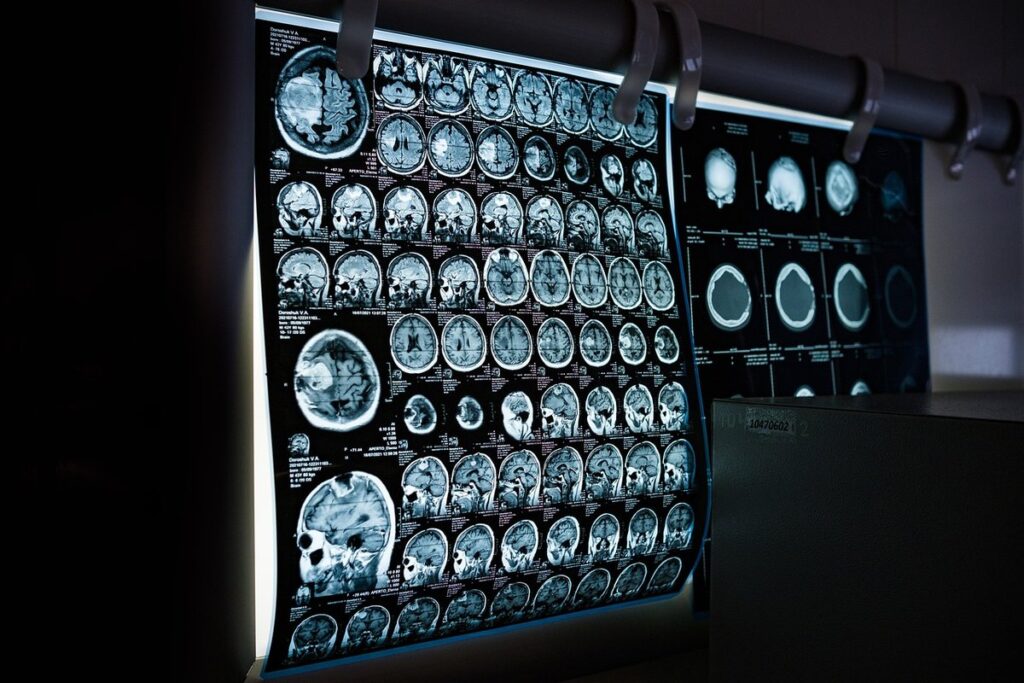
Diagnosing dementia often involves a combination of clinical assessments and advanced medical imaging techniques, including Magnetic Resonance Imaging (MRI). An MRI scan can provide valuable insights into brain structure and function, helping doctors identify potential causes of cognitive decline. You may have received an MRI report indicating changes in brain volume or white matter lesions, but what does it all mean? Understanding how to interpret these results is crucial for patients, families, and caregivers navigating the complexities of dementia diagnosis and treatment. In this article, we’ll explore the role of MRI scans in diagnosing dementia, including common types of scans, how to read MRI reports, and advanced techniques for monitoring treatment response. By the end, you’ll have a better grasp on how MRI results contribute to an accurate dementia diagnosis and be empowered to ask informed questions about your care.
What is Dementia and How Does It Relate to Brain Imaging?
Understanding dementia begins with understanding how it affects brain function, which is where brain imaging comes into play. We’ll break down what happens in the brain when dementia occurs and how that shows up on MRI scans.
Defining Dementia and Its Types
Dementia is a complex condition characterized by cognitive decline, affecting memory, thinking, and behavior. To better understand its diagnosis and management through MRI results, it’s essential to recognize the different types of dementia.
Alzheimer’s disease is the most common type, accounting for 60-80% of cases. It progresses gradually, with symptoms typically appearing after age 65. Vascular dementia, on the other hand, occurs due to reduced blood flow to the brain, often as a result of a series of small strokes or changes in blood vessels.
Frontotemporal dementia affects the front and temporal lobes, leading to changes in personality, language, and motor skills. Lewy body dementia combines cognitive decline with visual hallucinations and fluctuations in alertness. Other types include mixed dementia, where both Alzheimer’s and vascular factors contribute to symptoms, and rare conditions like Creutzfeldt-Jakob disease.
Understanding the specific type of dementia can help guide treatment and prognosis. A correct diagnosis is crucial for developing a personalized care plan. If you suspect a loved one may be experiencing cognitive decline, consult with a healthcare professional to discuss potential causes and next steps. They will typically conduct a comprehensive evaluation, including medical history, physical examination, and potentially an MRI scan to rule out underlying conditions affecting brain function.
The Role of MRI in Diagnosing Dementia
MRI plays a crucial role in diagnosing dementia by providing detailed images of brain structures. Healthcare professionals use various types of MRI scans to identify potential causes of cognitive decline. Functional MRI (fMRI) helps study brain activity, while diffusion tensor imaging (DTI) is used for tractography and white matter analysis.
Magnetic Resonance Angiography (MRA) is another type of MRI scan that helps detect vascular dementia, a condition caused by reduced blood flow to the brain. MRA uses magnetic fields and radio waves to visualize blood vessels in the brain.
The choice of MRI sequence depends on the specific needs of each patient. For example, Fluid-Attenuated Inversion Recovery (FLAIR) is often used to detect lesions that may be indicative of dementia. T1-weighted images are also commonly used to identify atrophy and other structural changes associated with dementia.
A healthcare professional will typically review MRI results alongside clinical data to establish a diagnosis. It’s essential for patients to discuss any concerns or questions they have about their MRI results with their doctor, as this can help inform treatment decisions and ensure the best possible outcomes.
How MRI Scans Work
To understand what your MRI scan results mean for a dementia diagnosis, it’s essential to grasp how these scans work and what they can reveal about brain changes. Let’s break down the basics of MRI technology.
Principles of MRI Technology
Magnetic fields are the foundation of MRI technology. In an MRI machine, a strong magnetic field is generated by superconducting magnets. This field is incredibly powerful – about 10,000 times stronger than the Earth’s magnetic field – and it’s what aligns the hydrogen protons in the brain. Hydrogen protons are abundant in water molecules within the body.
When exposed to this magnetic field, the aligned hydrogen protons absorb radio waves of a specific frequency. These radio waves are emitted by coils surrounding the magnet, and they induce an electrical signal that’s detected by the MRI machine. The strength and pattern of these signals vary depending on the orientation of the protons relative to the magnetic field.
Signal processing algorithms then convert this information into detailed images of the brain. This process is called spin echo formation, where the returning signal is amplified and processed multiple times to create high-resolution images. Understanding how MRI technology works is essential for accurately interpreting results and identifying abnormalities indicative of dementia.
Types of MRI Sequences Used in Brain Imaging
MRI sequences play a crucial role in producing detailed images of brain structures during an MRI scan. There are several types of sequences used in brain imaging, each with its unique characteristics and contributions to diagnosing dementia.
T1-weighted images provide excellent anatomical detail and are often used as a baseline for comparison with other sequences. They highlight the boundaries between different brain tissues and are particularly useful for visualizing grey matter. T2-weighted images, on the other hand, are more sensitive to changes in water content and are commonly used to detect abnormalities such as inflammation or edema.
FLAIR (Fluid-Attenuated Inversion Recovery) imaging is another essential sequence that helps identify lesions or areas of damage in the brain. By suppressing signal from fluids, FLAIR images make it easier to spot subtle changes in brain tissue that may be indicative of dementia.
In practice, radiologists often use a combination of these sequences to gain a comprehensive understanding of brain structure and function. For example, T1-weighted images might be used to identify atrophy or shrinkage of brain regions, while FLAIR imaging is employed to detect areas of damage or inflammation.
Interpreting MRI Results for Dementia
When reviewing your loved one’s MRI results, understanding what they mean can be a daunting task. This section will break down how to accurately interpret MRI findings related to dementia diagnosis.
Identifying Key Brain Structures on an MRI Scan
When interpreting an MRI scan for dementia, it’s essential to identify key brain structures that are commonly affected by the condition. The hippocampus, a seahorse-shaped structure crucial for memory formation, is often one of the first areas to show signs of damage. Located within the temporal lobe, the hippocampus plays a critical role in encoding and consolidating new memories.
Changes in the hippocampus can be an early indicator of dementia, particularly Alzheimer’s disease. Shrinkage or atrophy in this region can lead to difficulties in forming new memories, while sparing existing ones. The temporal lobe itself, which includes the hippocampus, is also frequently affected by dementia. Atrophy in this area can cause problems with language, mood regulation, and spatial awareness.
Other key brain structures visible on an MRI scan include the parietal lobe, involved in processing sensory information from various parts of the body, and the posterior cingulate cortex, which plays a role in attention and memory retrieval. Changes in these areas are often associated with dementia, particularly Alzheimer’s disease. Identifying these structural changes can help radiologists and clinicians make an accurate diagnosis and develop an effective treatment plan.
Recognizing Abnormalities Indicative of Dementia
When examining MRI results for dementia, several abnormalities often come to light. One common finding is white matter lesions, which appear as bright spots on the images. These lesions are typically a sign of small vessel disease and can be associated with vascular dementia. They may also indicate other conditions, such as multiple sclerosis or brain injuries.
Cortical atrophy, another notable abnormality, refers to the shrinking of brain tissue in the cerebral cortex. This can lead to a decrease in cognitive function and is often seen in Alzheimer’s disease patients. Ventricular enlargement, where the fluid-filled spaces within the brain become larger, may also be present. In some cases, ventricles may even appear as a dark spot on MRI scans.
It’s essential for healthcare professionals to carefully assess these abnormalities and consider their context when diagnosing dementia. While they can provide valuable clues, no single finding is definitive on its own. A comprehensive evaluation incorporating medical history, physical examination, and other diagnostic tools is crucial for an accurate diagnosis.
The Limitations of MRI in Diagnosing Dementia
While MRIs can provide valuable insights into brain health, they’re not a definitive diagnostic tool for dementia. This section explores the limitations and challenges of relying on MRI results alone to diagnose this complex condition.
Factors Affecting MRI Accuracy
MRI accuracy is influenced by several factors, which can be broadly categorized into image quality, scanner type, and patient variables. Image quality refers to the sharpness and clarity of the MRI images. A high-quality image ensures that even subtle changes in brain structure are visible. Factors affecting image quality include the strength of the magnetic field, the radiofrequency coil used, and the sequence parameters. For instance, a stronger magnetic field can produce higher-resolution images.
Scanner type is another critical factor affecting MRI accuracy. Different scanner models have varying levels of resolution and sensitivity to specific tissues or conditions. Some scanners are designed for specific applications, such as functional MRI (fMRI) or diffusion tensor imaging (DTI). When choosing an MRI machine for dementia diagnosis, consider the availability of high-resolution sequences and the scanner’s ability to produce detailed images of small brain structures.
Patient variables also play a significant role in determining MRI accuracy. Age is one such factor, as older adults may have reduced signal-to-noise ratio due to metal implants or other age-related changes. Additionally, medical history can impact image quality, with certain conditions, like claustrophobia or pacemakers, requiring special precautions during the scan.
Other Diagnostic Tools and Their Relationship to MRI Results
When diagnosing dementia, healthcare professionals often rely on a multi-faceted approach that incorporates various diagnostic tools beyond MRI scans. Laboratory tests, such as blood work and biomarker analysis, can provide valuable information about the presence of underlying conditions like Alzheimer’s disease or other neurodegenerative disorders.
For instance, a blood test may reveal elevated levels of beta-amyloid, a protein associated with Alzheimer’s, which can contradict MRI findings that indicate normal brain structure. Conversely, a clinical assessment may highlight cognitive decline and behavioral changes that support an MRI diagnosis of dementia.
Other diagnostic tools, such as electroencephalography (EEG), functional magnetic resonance imaging (fMRI), and positron emission tomography (PET) scans, can also be used to complement or contradict MRI results. EEG measures electrical activity in the brain, while fMRI examines brain function by detecting changes in blood flow. PET scans use small amounts of radioactive material to visualize brain metabolism.
These tests are not mutually exclusive, and their combination can provide a more comprehensive understanding of dementia’s progression and impact on brain health. A well-integrated diagnostic approach ensures that healthcare professionals consider all relevant information when making a diagnosis and developing treatment plans.
Advanced Imaging Techniques for Dementia Diagnosis
As we explore the complex world of dementia diagnosis, let’s take a closer look at how advanced imaging techniques can help doctors identify and treat the condition more effectively.
Functional MRI (fMRI) and Its Role in Studying Brain Activity
Functional MRI (fMRI) measures brain activity by detecting changes in blood flow to specific areas of the brain. In the context of dementia research, fMRI is used to study the neural correlates of cognitive decline and investigate the neural mechanisms underlying various dementias. For instance, researchers may use fMRI to compare brain activity patterns between individuals with Alzheimer’s disease and healthy controls during tasks such as memory encoding or word retrieval.
The benefits of using fMRI in dementia research include its ability to provide detailed information about brain function and connectivity. This can help researchers identify biomarkers for early diagnosis and develop targeted treatments. However, there are also limitations to consider. For example, fMRI is sensitive to motion artifacts, which can be a problem in individuals with advanced dementia who may have difficulty remaining still during the scanning process.
To mitigate this issue, researchers often use techniques such as task-based fMRI or resting-state fMRI, which involve simpler tasks or no explicit instructions at all. By understanding the strengths and limitations of fMRI in studying brain activity, researchers can design more effective studies that ultimately improve our understanding of dementia and inform treatment development.
Diffusion Tensor Imaging (DTI) for Tractography and White Matter Analysis
DTI produces detailed maps of white matter tracts, allowing researchers to assess their integrity. This is particularly relevant for dementia diagnosis, as white matter degeneration is a common feature across various types of dementia. DTI can identify areas where fiber tracts are damaged or disrupted, which may correlate with cognitive decline.
For example, studies have used DTI to investigate the relationship between white matter lesions and dementia severity. By analyzing DTI data, researchers can gain insights into how these lesions affect normal brain function. This information can be crucial for developing targeted treatments that address specific aspects of dementia pathology.
DTI also enables the creation of detailed tractography maps, which can help clinicians understand the anatomical connectivity of affected regions. This can inform diagnosis and treatment planning by highlighting areas where interventions may have a significant impact.
In practice, DTI results are often compared to other imaging modalities, such as structural MRI or functional MRI, to gain a more comprehensive understanding of brain function in individuals with dementia. By combining these data types, researchers and clinicians can develop a more nuanced picture of the complex relationships between white matter degeneration, cognitive decline, and dementia pathology.
Managing Dementia with MRI-Guided Therapy
As you consider treatment options for a loved one, learning how MRI-guided therapy can manage dementia is crucial to making informed decisions. This approach offers promising results in slowing disease progression and improving quality of life.
The Role of MRI in Monitoring Treatment Response
Regular follow-up MRI scans are essential for monitoring treatment efficacy in patients with dementia. These scans can detect subtle changes in brain structure and function, allowing clinicians to adjust therapeutic plans accordingly.
For instance, if a patient is receiving cholinesterase inhibitor therapy, regular MRI scans can help assess whether the medication is effectively reducing atrophy rates or slowing cognitive decline. Conversely, if the treatment appears ineffective, clinicians may opt for alternative medications or complementary therapies.
In addition to monitoring response to treatment, ongoing imaging evaluation can also help identify potential side effects of medications on brain function. For example, certain antipsychotic medications have been linked to increased risk of cerebrovascular events in patients with dementia; MRI scans can detect such changes and inform adjustments to the patient’s medication regimen.
To make the most of this monitoring process, clinicians should work closely with radiologists to review and interpret MRI results regularly. By doing so, they can refine treatment plans, improve patient outcomes, and optimize the use of limited healthcare resources.
Emerging Therapies and Their Potential Impact on MRI Results
New treatments, such as immunotherapy or gene therapies, aim to slow down or halt disease progression. These emerging therapies have sparked interest in their potential impact on MRI results, particularly in monitoring treatment efficacy and disease progression.
Immunotherapy, for instance, has shown promise in targeting specific proteins associated with dementia. If successful, these treatments could lead to changes in brain atrophy patterns visible on MRI scans. However, the initial effects might be subtle, requiring longer-term follow-up imaging studies to detect significant differences.
Gene therapies also hold potential in modifying disease-causing genes. MRI can provide valuable insights into the efficacy of such interventions by monitoring changes in brain structure and function over time. For example, researchers have used MRI to track the growth of new neurons in response to gene therapy.
As these emerging therapies continue to evolve, future research will likely focus on understanding their long-term effects on MRI results. This could involve developing more sensitive imaging protocols or exploring novel biomarkers that better capture treatment responses. By closely monitoring these developments, healthcare professionals and researchers can refine their approaches to managing dementia with MRI-guided therapy.
Frequently Asked Questions
Can I get an MRI scan on an emergency basis to diagnose dementia?
Yes, in cases of acute cognitive decline or suspected stroke, healthcare providers may order an urgent MRI scan. However, it’s essential to note that these scans are typically performed in a hospital setting and may require immediate attention from radiologists and neurologists.
What if my MRI results show some abnormalities but the doctor says they’re not indicative of dementia? Should I be concerned?
Yes, it’s natural to have questions and concerns. Even if your doctor doesn’t suspect dementia based on your MRI results, other conditions or factors might be contributing to your symptoms. Discuss any concerns you have with your healthcare provider, who can help interpret the results in context.
How long does it take for changes in brain structure visible on an MRI scan to indicate dementia progression?
The time frame varies depending on individual cases and the specific type of dementia. However, research suggests that early changes in brain structure, detectable through advanced imaging techniques like diffusion tensor imaging (DTI), can occur several years before clinical symptoms manifest.
Can I use MRI results as a benchmark for monitoring treatment efficacy in patients with dementia?
Yes, regular follow-up MRI scans can help healthcare providers assess treatment response and adjust therapeutic plans accordingly. However, it’s crucial to understand that MRI results should be interpreted in conjunction with other diagnostic tools and clinical assessments.
What if my doctor recommends an MRI scan but I’m claustrophobic? Are there alternative options available?
Yes, there are alternative MRI machines designed for patients who experience anxiety or claustrophobia. Open-bore or wide-bore MRI scanners can provide a more comfortable environment during the scanning process. Discuss your concerns with your healthcare provider to explore these options and find the best solution for you.