
Dementia diagnosis is often a complex process, relying heavily on accurate detection of brain changes. One crucial tool in this diagnostic journey is the CT scan, which has become an essential component of dementia diagnosis due to its ability to provide detailed images of brain structure and function. While traditional methods like MRI are also used, CT scans offer a unique set of benefits that make them particularly well-suited for detecting early signs of dementia. By understanding how CT scans work and what they can reveal about the brain, medical professionals can make more informed decisions about patient care. In this article, you’ll learn how CT scans play a vital role in diagnosing dementia, including principles of technology, detecting brain changes, and interpreting results to provide accurate diagnoses.
Understanding Dementia and the Role of Imaging
Dementia is a complex condition, and understanding its underlying causes and progression can be a challenge. This section will help you grasp the basics of dementia and how imaging plays a crucial role in diagnosis.
The Importance of Early Diagnosis
Early diagnosis of dementia is crucial for effective management. Timely detection enables healthcare providers to initiate treatment and slow disease progression. Imaging plays a pivotal role in this process by providing valuable insights into brain structure and function.
Delays in diagnosis can have significant consequences, including accelerated cognitive decline and reduced quality of life. In contrast, early intervention can improve outcomes and enhance patient care. For instance, medications such as cholinesterase inhibitors are most effective when administered during the early stages of dementia.
Imaging modalities like CT scans help identify changes in brain structure associated with dementia. These changes include atrophy, particularly in areas responsible for memory and cognitive function. By detecting these alterations, healthcare providers can initiate appropriate treatment plans tailored to each patient’s needs.
Prompt diagnosis also enables caregivers to develop informed care strategies, ensuring the best possible support for patients. Ultimately, early detection through imaging modalities like CT scans is essential for maximizing dementia management outcomes.
Overview of Imaging Modalities
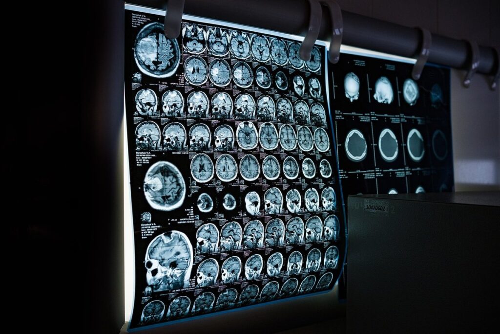
CT scans are one of several imaging modalities used to diagnose dementia. They produce detailed cross-sectional images of brain tissue, allowing healthcare professionals to identify signs of disease progression and structural changes. Magnetic Resonance Imaging (MRI) is another widely used modality that provides high-resolution images of the brain’s internal structures. However, MRI scans are generally more sensitive to soft tissue differentiation than CT scans.
Positron Emission Tomography (PET) scans also play a crucial role in dementia diagnosis. PET uses small amounts of radioactive tracers to visualize changes in brain metabolism and blood flow. This modality is particularly useful for detecting the spread of neurodegenerative diseases, such as Alzheimer’s. Each imaging modality has its strengths and limitations.
CT scans are often preferred due to their ability to quickly produce detailed images of bone and soft tissue structures. However, PET scans offer more sensitive measurements of brain metabolism. MRI scans provide high-resolution images but may require longer scanning times. When selecting an imaging modality for dementia diagnosis, healthcare professionals consider factors such as the patient’s age, medical history, and symptoms. This multi-modal approach helps ensure accurate diagnosis and effective treatment planning.
How CT Scans Aid in Dementia Diagnosis
CT scans have become a crucial tool in diagnosing dementia, helping doctors identify physical changes in the brain that can confirm a diagnosis. Let’s explore how these scans aid in detecting the signs of this complex condition.
Principles of CT Scan Technology
CT scans use a process called X-ray computed tomography to generate detailed cross-sectional images of the brain. This technology is based on the principle that different tissues absorb varying levels of X-rays, allowing for distinction between them. The scanner emits a beam of X-rays that passes through the body and strikes a detector array on the opposite side.
The detector measures the intensity of the X-rays as they exit the body, providing data used to reconstruct images. Image reconstruction is achieved by applying mathematical algorithms to the raw data, which involves solving a set of equations to estimate the distribution of X-ray absorption coefficients within the scanned volume.
Key factors in image quality include slice thickness and resolution. A thinner slice provides more detailed images but may compromise on spatial resolution due to reduced signal-to-noise ratio. Typical slice thickness ranges from 1-5 mm, depending on the scanner model and clinical requirements. In practice, optimal settings for dementia diagnosis typically involve a balance between these competing factors.
Detecting Changes in Brain Structure
When analyzing CT scan images for dementia diagnosis, radiologists look for specific changes in brain structure. One key indicator is the presence of atrophy, which refers to a reduction in brain tissue volume. This can be seen as a shrinkage of the outer layers of the brain or a thinning of the cerebral cortex. Atrophy is particularly noticeable in areas responsible for memory and cognitive function, such as the hippocampus and temporal lobes.
Another sign of dementia-related changes on CT scans is the accumulation of cerebrospinal fluid (CSF) in certain regions. This can be seen as a bright spot or a “whiteout” effect on the scan image. The increased CSF can indicate damage to brain tissue, which may be indicative of neurodegenerative disease.
In addition to these changes, radiologists also examine the ventricles and sulci for enlargement or widening. These structures become more pronounced as atrophy progresses, allowing radiologists to identify potential signs of dementia on CT scans.
CT Scan vs. Other Imaging Modalities
Compared to other imaging modalities, a CT scan stands out for its ability to detect specific biomarkers associated with dementia in the brain. Let’s examine how it measures up against MRI and PET scans.
Comparison with MRI Scans
When it comes to diagnosing dementia, both CT scans and MRI scans are used as imaging modalities. However, they have distinct advantages and limitations. One key difference is the level of detail each scan provides. MRI scans offer high-resolution images that can detect subtle changes in brain structure, making them ideal for identifying conditions like multiple sclerosis or stroke. In contrast, CT scans provide faster image acquisition and are better suited for detecting larger abnormalities, such as tumors or fractures.
A study published in the Journal of Alzheimer’s Disease found that while both modalities were effective in detecting dementia, MRI scans were more sensitive than CT scans in identifying early-stage disease. However, this comes at a cost: MRI scans are typically longer and may require more patient preparation. For instance, metal-free clothing and implants must be removed before the scan to prevent artifacts.
Ultimately, the choice between CT and MRI scans depends on the specific clinical scenario. A radiologist’s expertise will guide this decision, taking into account factors like available equipment, patient condition, and desired diagnostic outcomes.
PET Scans: A Different Approach
PET scans use a different approach to image brain activity and metabolic changes associated with dementia. Unlike CT scans, which primarily focus on structural changes, PET scans measure the distribution of glucose throughout the brain. This allows for the detection of areas where glucose uptake is altered, often indicative of dementia-related pathology.
In practice, PET scans may be preferred when a more detailed assessment of brain function is required, such as in cases where CT scans have produced inconclusive results or when additional diagnostic information is needed to inform treatment decisions. For example, in some studies, PET scans using the tracer fluorodeoxyglucose (FDG) have been shown to detect changes in brain glucose metabolism associated with Alzheimer’s disease.
When comparing PET and CT scans for dementia diagnosis, it’s essential to consider the trade-offs between each modality. While CT scans are generally faster and more widely available, PET scans offer a unique window into brain function and may provide valuable additional information in specific cases.
Factors Influencing CT Scan Accuracy
Accurate CT scan results are crucial for a reliable dementia diagnosis, so let’s examine how various factors can impact their accuracy. These influencing elements include patient preparation and medical history.
Patient Selection Criteria
Patient selection is a critical step in determining whether a CT scan is suitable for diagnosing dementia. Age is one key factor to consider: patients typically need to be at least 18 years old, but older adults may require additional considerations due to comorbidities or reduced cognitive function.
A patient’s medical history can also impact their suitability for a CT scan. For instance, those with a history of kidney disease or contrast allergy should not undergo CT scans using iodine-based contrast agents. Additionally, patients with certain metallic implants or devices, such as pacemakers, may need to be referred to an MRI instead.
Cognitive status is another crucial factor in patient selection. Patients with severe cognitive impairment may not be able to follow instructions or remain still during the scan, which can compromise image quality. In these cases, alternative imaging modalities like PET scans may be more suitable. Healthcare providers should carefully evaluate each patient’s individual circumstances and discuss their suitability for a CT scan before proceeding.
When evaluating patients, healthcare providers often consider the following:
Image Acquisition and Analysis Techniques
To ensure accurate CT scan diagnoses, it’s crucial to employ proper image acquisition techniques. This involves optimizing scanner settings for each patient, taking into account factors such as age, weight, and medical history. For example, patients with a higher body mass index may require adjustments to beam energy or scanning time to compensate for increased attenuation.
A well-trained technologist should carefully position the patient within the scanner’s field of view to minimize motion artifacts. The use of breath-hold instructions can also help reduce respiratory motion effects on image quality. Furthermore, selecting the correct slice thickness and reconstruction algorithm is vital for maintaining image resolution while minimizing radiation exposure.
When it comes to analysis techniques, radiologists should follow a standardized workflow for reviewing CT scan images. This includes evaluating brain structure changes, such as atrophy or ventricular enlargement, in conjunction with assessing cortical thickness and white matter abnormalities. Attention should be given to the protocol’s specific requirements for identifying dementia-related features.
The Role of AI and Deep Learning in CT Scan Interpretation
Recent advancements in artificial intelligence and deep learning have significantly improved the accuracy and efficiency of CT scan interpretation, particularly in diagnosing dementia.
These innovative technologies enable radiologists to identify subtle changes in brain structure that may be indicative of cognitive decline.
Current Applications and Limitations
AI and deep learning algorithms have been successfully integrated into various medical imaging applications, including CT scan interpretation for dementia diagnosis. Currently, these technologies are mainly used to automate image analysis tasks, such as segmenting brain structures or detecting white matter hyperintensities. For instance, a study published in the Journal of Alzheimer’s Disease demonstrated that a deep learning-based algorithm achieved high accuracy in identifying patients with Alzheimer’s disease using CT scans.
However, there are still limitations to the current applications of AI and deep learning in CT scan interpretation for dementia diagnosis. One major challenge is data quality and availability – most existing datasets are small and biased towards specific populations, which can affect model generalizability. Furthermore, the lack of standardization in image acquisition protocols and analysis techniques hinders the development of robust and transferable models. To overcome these limitations, researchers and clinicians must work together to create more comprehensive and diverse datasets, as well as establish best practices for image acquisition and analysis.
Potential Future Developments
Advances in AI and deep learning will likely lead to more sophisticated image analysis techniques, allowing for better detection of subtle changes in brain structure associated with dementia. Future developments may also incorporate multimodal imaging, combining CT scans with other modalities like MRI or PET scans to provide a more comprehensive understanding of brain health.
Researchers are exploring the use of transfer learning and domain adaptation to improve AI models’ performance on diverse datasets. This could enable better generalizability across different patient populations and scanning protocols. Additionally, advancements in data augmentation techniques may help mitigate the limitations of small dataset sizes, common in medical imaging research.
Another promising area is the integration of clinical knowledge into AI decision-making processes. This could involve developing hybrid models that combine the strengths of machine learning with expert medical knowledge. By doing so, clinicians can leverage the accuracy of AI while still benefiting from human expertise and contextual understanding.
Future studies may focus on the development of explainable AI (XAI) methods for CT scan interpretation. XAI techniques would provide insights into how AI models arrive at their conclusions, enabling clinicians to better understand and trust AI-driven diagnoses. This could lead to more accurate and reliable dementia diagnoses through enhanced collaboration between humans and machines.
Interpreting CT Scan Results: A Step-by-Step Guide
Understanding your CT scan results can be overwhelming, so we’ll break down the key points to look for in a step-by-step guide. This will help you navigate what each finding means for your loved one’s diagnosis and treatment.
Understanding the Report and Recommendations
When you receive a CT scan report for dementia diagnosis, it’s essential to understand what the results mean and how they relate to your specific situation. Begin by reviewing the report’s summary section, which should provide an overview of the findings and recommendations.
Look for key terms such as “abnormalities” or “changes in brain structure,” which indicate potential issues that may be related to dementia. The report may also mention specific areas of the brain affected, such as the hippocampus or temporal lobe, and how these changes correspond to your symptoms.
The recommendations section should outline next steps for further evaluation or treatment. This may include additional imaging tests, neurological exams, or consultations with specialists. Pay attention to any notes or comments from the radiologist, which can provide valuable context for interpreting the results.
When reviewing the report, keep in mind that CT scans are often used as a screening tool to identify potential dementia cases. A positive result doesn’t necessarily mean a diagnosis of dementia, but rather indicates further evaluation is needed.
Communicating with Healthcare Providers
When communicating CT scan results and recommendations to healthcare providers, it’s essential to be clear and concise. Start by reviewing the report and identifying key findings, such as changes in brain structure or atrophy patterns. Make sure to understand the radiologist’s interpretation and ask questions if you’re unsure about any aspect of the report.
Be prepared to discuss your medical history, including previous diagnoses and treatments. Share relevant information about your symptoms, such as memory loss, confusion, or difficulty with daily activities. This context is crucial for healthcare providers to make informed decisions about your care.
When discussing CT scan results, focus on the specific findings rather than generalizing about your condition. For example, instead of saying “I have dementia,” say “The CT scan indicates significant atrophy in the temporal lobe, which may be indicative of Alzheimer’s disease.” This helps healthcare providers understand the nuances of your diagnosis and develop a tailored treatment plan.
When communicating with healthcare providers, consider bringing a family member or caregiver for support. It can also help to keep a record of your conversations, including dates, times, and details discussed.
Conclusion: The Future of Dementia Diagnosis
Now that we’ve covered the process and benefits of CT scan dementia diagnosis, let’s consider what advancements might shape its future. Emerging technologies hold promise for improved accuracy and patient outcomes.
Advancements in Imaging Technology
Advances in imaging technology are poised to revolutionize dementia diagnosis and management. One notable development is the increasing use of artificial intelligence (AI) to enhance image analysis and interpretation. AI algorithms can quickly identify subtle changes in brain structure, which may not be apparent to human radiologists. For instance, researchers have developed AI-powered tools that can detect early signs of Alzheimer’s disease, such as atrophy in specific brain regions.
Another promising area is the development of advanced imaging modalities, such as high-field MRI and positron emission tomography (PET) scans. These technologies offer improved resolution and sensitivity, enabling clinicians to identify subtle changes in brain function and structure. Additionally, advancements in image acquisition techniques, such as diffusion tensor imaging, are providing new insights into the underlying pathology of dementia.
The integration of these advanced technologies with CT scans is also on the horizon. By combining CT’s ability to visualize large areas of the brain with AI-powered analysis, clinicians may be able to detect dementia earlier and more accurately than ever before.
Emerging Trends and Research Directions
Researchers are actively exploring new applications of CT scan technology for dementia diagnosis. One promising area is the use of artificial intelligence (AI) and machine learning algorithms to enhance image analysis and improve diagnostic accuracy. These tools can help identify subtle changes in brain structure that may be indicative of dementia, even before symptoms appear.
Another emerging trend is the integration of multimodal imaging techniques, which combine CT scans with other modalities such as MRI or PET scans. This approach allows for a more comprehensive understanding of brain function and anatomy, potentially leading to earlier and more accurate diagnoses.
Additionally, researchers are investigating the use of advanced image processing techniques, such as diffusion tensor imaging (DTI) and magnetic resonance elastography (MRE), to better visualize white matter tracts and detect subtle changes in brain tissue. These developments hold great promise for improving CT scan-based dementia diagnosis and may become increasingly important in clinical practice in the future.
Some studies are also exploring the use of CT scans to monitor disease progression and response to treatment, which could revolutionize the way clinicians manage dementia patients. While these advancements are still in their early stages, they demonstrate the rapidly evolving nature of this field and highlight the potential for significant improvements in dementia diagnosis using CT scan technology.
Frequently Asked Questions
How Long Does It Take to Get CT Scan Results for Dementia Diagnosis?
Getting CT scan results can take anywhere from a few hours to several days, depending on the facility’s workload and the complexity of the images. In most cases, your healthcare provider will receive a preliminary report within 24-48 hours, with a final diagnosis typically available within 1-3 business days.
Can AI-Enhanced CT Scans Be Used for Dementia Diagnosis in Rural or Remote Areas?
Yes, AI-enhanced CT scans can be used for dementia diagnosis in rural or remote areas. These technologies enable healthcare providers to interpret images more accurately and efficiently, reducing the need for specialized facilities or on-site experts. This can help bridge the gap in accessing quality care in underserved regions.
What if My Healthcare Provider Is Unfamiliar with Interpreting CT Scan Results for Dementia Diagnosis?
If your healthcare provider is unfamiliar with interpreting CT scan results for dementia diagnosis, they may recommend consulting a specialist or seeking a second opinion from a facility with more experience in this area. This ensures that you receive accurate and informed care.
Can I Get a Second Opinion on My CT Scan Results Using the Same Modality (e.g., Another CT Scan)?
Yes, you can get a second opinion on your CT scan results using the same modality. Many facilities offer this service, allowing you to have your images reevaluated by an independent expert. This can provide peace of mind and help confirm or rule out a diagnosis.
What if My CT Scan Results Indicate Changes in Brain Structure but Don’t Provide a Clear Diagnosis?
If your CT scan results indicate changes in brain structure but don’t provide a clear diagnosis, your healthcare provider may recommend additional imaging tests (e.g., MRI or PET scans) to gather more information. They may also discuss the implications of these findings and potential next steps for further evaluation and management.