
Accurate early detection is key to managing dementia effectively. While memory loss and cognitive decline can be distressing, modern medical advancements have made significant strides in identifying these conditions through magnetic resonance imaging (MRI). This non-invasive diagnostic tool helps healthcare professionals visualize brain changes associated with dementia, such as atrophy or vascular damage. By leveraging MRI scans, doctors can develop personalized management plans to slow disease progression and improve quality of life for patients. In this article, we’ll explore the crucial role MRI plays in early dementia diagnosis and how it enables timely intervention. We’ll delve into the specifics of how MRI technology works and its benefits for patients, as well as discuss the importance of accurate diagnosis in improving outcomes.
What is Dementia and Why is Early Diagnosis Important?
Dementia is a complex condition that affects millions of people worldwide, and understanding its causes and symptoms is crucial for early detection. Let’s break down what dementia is and why getting an accurate diagnosis as soon as possible matters greatly.
Prevalence and Types of Dementia
Dementia is a complex neurodegenerative disorder characterized by cognitive decline and memory loss. According to the World Health Organization (WHO), approximately 50 million people worldwide suffer from dementia, with numbers expected to triple by 2050. Alzheimer’s disease accounts for the majority of dementia cases, affecting over 60% of individuals. Vascular dementia is the second most common type, resulting from reduced blood flow to the brain due to impaired blood vessels.
Lewy body dementia, a lesser-known variant, is caused by abnormal protein clumps called Lewy bodies in the brain. Each type of dementia has distinct symptoms and progression rates. For instance, Alzheimer’s disease typically follows a predictable pattern, with early stages marked by memory loss and difficulty learning new information. Vascular dementia often presents with abrupt cognitive decline following a stroke or transient ischemic attack.
Accurate diagnosis is essential for effective management and treatment planning. MRI plays a crucial role in identifying structural changes associated with various types of dementia. By understanding the different forms of dementia, healthcare professionals can tailor diagnostic approaches to individual patients’ needs, ensuring timely intervention and optimizing care outcomes.
Importance of Early Diagnosis and Intervention
Early diagnosis and intervention are crucial in dementia care. When symptoms first appear, individuals with dementia can still engage in daily activities, maintain their independence, and enjoy a better quality of life. Delaying diagnosis by just a few years can significantly impact this trajectory.
Caregivers also benefit from early detection. They often experience increased stress, emotional burden, and physical exhaustion when caring for loved ones with advanced dementia. Early intervention allows caregivers to receive support and respite services, reducing their burnout risk.
Early diagnosis may also slow disease progression by enabling timely initiation of effective treatments. Some medications can manage symptoms or even alter the course of certain dementias. In addition to medication, lifestyle modifications such as cognitive training, exercise, and social engagement have shown promise in slowing decline. While these interventions are not a guarantee against dementia’s eventual progression, they may delay its onset.
Studies suggest that early intervention can also reduce healthcare costs associated with advanced dementia care, which can be substantial.
The Role of MRI in Dementia Diagnosis
MRI plays a crucial role in dementia diagnosis, providing valuable insights into brain structure and function that can aid in early detection and treatment planning. We’ll take a closer look at how MRI helps doctors diagnose dementia accurately.
Introduction to MRI Technology and Its Application in Neurodegenerative Disorders
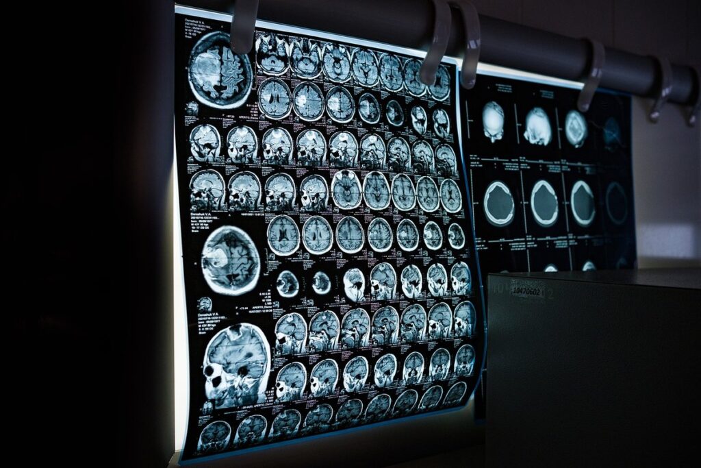
Magnetic resonance imaging (MRI) technology is based on the principle of nuclear magnetic resonance. This involves aligning hydrogen atoms within the body and then measuring the signals they emit when exposed to a strong magnetic field and radiofrequency pulses. The resulting images provide detailed information about tissue structure, water content, and blood flow. In neurodegenerative disorders like dementia, MRI can detect changes in brain anatomy, such as atrophy or lesions, which are indicative of disease progression.
MRI has become an essential diagnostic tool for neurodegenerative diseases due to its non-invasive nature and high spatial resolution. Its application in neurodegenerative disorders involves using different sequences and protocols tailored to specific conditions. For example, T1-weighted images can highlight gray matter atrophy, while FLAIR (Fluid-Attenuated Inversion Recovery) sequences are sensitive to white matter lesions.
The MRI’s ability to assess subtle changes in brain tissue makes it an invaluable tool for tracking disease progression and monitoring treatment efficacy. By identifying early signs of neurodegeneration, clinicians can initiate timely interventions, potentially slowing down or halting disease progression. This highlights the critical role MRI plays in dementia diagnosis and management, particularly when used in conjunction with other diagnostic tools and clinical assessments.
MRI Findings Associated with Dementia
Hippocampal atrophy is a hallmark MRI finding associated with dementia. This refers to the shrinkage of the hippocampus, a critical brain region involved in memory formation and consolidation. Studies have consistently shown that individuals with Alzheimer’s disease exhibit significant hippocampal volume loss, particularly in the posterior hippocampus. Ventricular enlargement is another common MRI finding in people with dementia. The ventricles are fluid-filled spaces within the brain that surround the cerebral cortex, and their enlargement can be indicative of neurodegenerative processes.
White matter hyperintensities (WMH) are also a common feature on MRI scans of individuals with dementia. WMH are areas where the brain’s white matter appears bright due to inflammation or demyelination. They can be seen in various locations, including the periventricular and deep white matter regions. The presence and severity of WMH have been linked to cognitive decline and an increased risk of dementia.
These MRI findings can provide valuable information about the underlying pathophysiology of dementia and can help clinicians make a more accurate diagnosis. For example, hippocampal atrophy may suggest Alzheimer’s disease, while WMH could indicate vascular dementia or other forms of cognitive impairment.
MRI as a Diagnostic Tool for Dementia
MRI technology plays a vital role in diagnosing dementia by providing detailed images of brain structure and function, helping doctors pinpoint potential causes. We’ll examine how MRI is used to support an early diagnosis.
How MRI Helps Diagnose Dementia
MRI helps diagnose dementia by providing a detailed view of brain structure and function. In atypical presentations, where symptoms don’t fit typical Alzheimer’s disease patterns, MRI can identify unusual features such as cortical thinning or hippocampal atrophy. This is particularly useful for diagnosing frontotemporal dementia (FTD) and other less common forms.
MRI also plays a crucial role in monitoring disease progression. Regular imaging sessions enable clinicians to track changes in brain volume, white matter lesions, and other markers of neurodegeneration. For example, studies have shown that annual MRI scans can detect a significant decline in hippocampal volume within a year of the initial scan.
By identifying these changes early on, healthcare professionals can adjust treatment plans accordingly, potentially improving patient outcomes. Moreover, MRI findings can be used to monitor the effectiveness of interventions, such as medications or cognitive training programs. For instance, if an MRI shows that certain areas of the brain are shrinking at a slower rate after initiating a treatment, it may indicate that the therapy is effective in slowing disease progression.
Comparison with Other Diagnostic Tools (e.g., CT, PET)
Compared to other diagnostic tools like CT and PET scans, MRI offers several advantages. One key difference is that MRI does not use ionizing radiation, which can be a concern for patients who require repeated scans or have pre-existing conditions. In contrast, CT scans use X-rays to produce images, while PET scans rely on radioactive tracers.
Another advantage of MRI is its ability to provide high-resolution images of the brain’s structure and function. This allows doctors to identify specific patterns of atrophy or degeneration associated with dementia, such as hippocampal shrinkage or white matter lesions. Additionally, MRI can detect changes in brain metabolism and connectivity that may not be visible on other types of scans.
However, MRI also has some limitations when it comes to diagnosing dementia. For example, it may not always distinguish between different types of dementia or identify early stages of the disease. In some cases, MRI findings may need to be correlated with clinical symptoms and cognitive tests to confirm a diagnosis.
Advanced MRI Techniques in Dementia Research and Diagnosis
Advanced MRI techniques are revolutionizing dementia research by providing detailed images of brain changes, enabling researchers to better understand disease progression. These sophisticated methods hold great promise for early detection and improved diagnosis.
Diffusion Tensor Imaging (DTI) and Its Applications
DTI measures the diffusion of water molecules within brain tissue, providing insights into white matter integrity. This technique is based on the principle that water molecules move more easily through healthy tissues than those affected by disease or injury. In dementia research, DTI has been used to track changes in white matter tracts, which are often compromised early in the disease process.
Studies have shown that DTI can detect subtle alterations in brain connectivity even before noticeable cognitive decline. This makes it a valuable tool for identifying individuals at risk of developing dementia. For instance, researchers have found that patients with mild cognitive impairment (MCI) exhibit altered white matter tracts compared to healthy controls. These findings suggest that DTI may help clinicians identify early biomarkers of dementia.
DTI has also been used to study the effects of various interventions on brain health. For example, one study examined the impact of exercise on white matter integrity in individuals with MCI. Results indicated that regular physical activity improved white matter tracts, suggesting a potential therapeutic application for DTI-guided exercise programs. As research continues to advance our understanding of DTI’s role in dementia diagnosis and management, its applications are likely to expand further.
Functional MRI (fMRI) and Cognitive Mapping
Functional MRI (fMRI) uses magnetic fields and radio waves to measure changes in brain activity by detecting blood flow. In dementia research, fMRI is used to create cognitive maps – detailed diagrams of brain regions involved in various mental processes such as memory, language, or problem-solving. These maps help researchers understand how different areas of the brain interact and communicate with each other.
Studies have shown that individuals with Alzheimer’s disease exhibit altered patterns of brain activity compared to healthy controls. fMRI can detect these changes by highlighting specific brain regions that are overactive or underactive during certain tasks. This information can be used to identify potential biomarkers for dementia, which could lead to earlier diagnosis and more effective treatment.
fMRI cognitive maps can also help clinicians tailor interventions to an individual’s unique brain profile. For example, a patient with Alzheimer’s may show decreased activity in the hippocampus – a region critical for memory formation. An fMRI-based treatment plan might focus on exercises that stimulate this area, such as memory games or cognitive training programs.
By providing detailed insights into brain function and connectivity, fMRI and cognitive mapping offer valuable tools for understanding dementia and improving patient outcomes.
Challenges and Limitations of Using MRI for Dementia Diagnosis
While MRI has revolutionized dementia diagnosis, it’s not without its challenges. We’ll explore some of the key limitations that affect diagnostic accuracy and patient outcomes directly.
Radiation Safety Concerns and Alternative Options
Radiation safety concerns are a critical consideration when using MRI for dementia diagnosis. Ionizing radiation from MRI scans can pose health risks to patients and staff, particularly with repeated scans over time. However, it’s essential to note that the amounts of ionizing radiation used in MRI scans are generally low, comparable to those found in some types of computed tomography (CT) scans.
Non-ionizing imaging modalities have emerged as alternative options for dementia diagnosis. One such option is functional MRI (fMRI), which measures changes in blood flow to map brain function without using ionizing radiation. Another option is diffusion tensor imaging (DTI), which assesses the integrity of white matter tracts without exposing patients to radiation.
Researchers are also exploring the use of magnetic resonance spectroscopy (MRS) and magnetization transfer imaging (MTI) as non-ionizing alternatives for dementia diagnosis. These techniques can provide valuable information on brain metabolism, microstructure, and other factors relevant to dementia research. As these alternative options continue to develop, they may offer safer and more effective means of diagnosing dementia using MRI technology.
Limitations of Current Imaging Technologies and Future Directions
Current MRI technologies for dementia diagnosis have limitations in terms of resolution. Standard MRI scans often struggle to capture subtle changes in brain structure and function associated with early-stage dementia. This is particularly true for high-resolution imaging, which requires specialized equipment and expertise.
Another limitation lies in the sensitivity of contrast agents used in MRI scans. While gadolinium-based agents can enhance tissue contrast, they may not be effective in detecting certain types of neurodegeneration. Researchers have begun exploring alternative contrast agents and techniques to improve sensitivity and specificity.
To overcome these limitations, future directions focus on developing more advanced imaging protocols and technologies. Magnetic resonance elastography (MRE) and functional MRI (fMRI) are promising areas of research that aim to provide more accurate and detailed information about brain function and structure.
Developers also seek to optimize existing technologies by refining image acquisition parameters, improving data analysis algorithms, and integrating multimodal imaging approaches. By advancing these capabilities, researchers hope to create more effective tools for early dementia diagnosis and monitoring disease progression.
Best Practices for Using MRI in Dementia Diagnosis and Management
When using MRI scans to diagnose and manage dementia, there are several key practices that medical professionals should follow to ensure accurate results. These best practices involve careful consideration of imaging protocols and patient preparation.
Image Acquisition and Analysis Guidelines
To ensure accurate diagnosis and management of dementia, it’s essential to follow optimal image acquisition and analysis protocols. For MRI, a 1.5T or 3T scanner is typically used for brain imaging, with a minimum matrix size of 256 x 256 pixels and a slice thickness of 2-3 mm. Slice orientation should be parallel to the anterior commissure-posterior commissure (ACPC) line to minimize distortion.
A T1-weighted sequence with contrast agent is usually acquired first to provide anatomical details, followed by T2-weighted and fluid-attenuated inversion recovery (FLAIR) sequences for better visualization of lesions. Diffusion-weighted imaging (DWI) may also be used to assess white matter integrity.
When analyzing MRI images, radiologists should look for signs such as hippocampal atrophy, ventricular enlargement, and white matter hyperintensities. Bulleted guidelines for image interpretation include:
• Assessing the volume of the hippocampus and amygdala
• Evaluating the integrity of white matter tracts
• Identifying areas of cortical thinning or atrophy
Clinical Correlation and Integration with Other Diagnostic Tools
Accurate diagnosis and management of dementia require a comprehensive approach that integrates clinical correlation with other diagnostic tools. MRI findings are just one piece of the puzzle, and interpreting them requires consideration of the patient’s medical history, cognitive function, and other relevant factors. A thorough review of the patient’s chart, including laboratory results, medication lists, and prior imaging studies, is essential for clinicians to make informed decisions.
The American Academy of Neurology recommends that MRI findings be correlated with clinical data, including the patient’s Mini-Mental State Examination (MMSE) score and other neuropsychological assessments. This ensures that any abnormalities detected on an MRI are considered in the context of the patient’s overall clinical presentation. Clinicians should also consider alternative explanations for MRI findings, such as vascular disease or medication effects.
When integrating MRI with other diagnostic tools, clinicians can use a combination of quantitative measures, such as atrophy rates and white matter hyperintensities, to inform their diagnosis and management plan. For example, a patient with significant atrophy in the hippocampus may be diagnosed with Alzheimer’s disease, while a patient with prominent white matter hyperintensities may be diagnosed with vascular dementia.
Frequently Asked Questions
How Long Does an MRI Scan Take for Dementia Diagnosis?
MRI scans for dementia diagnosis typically take around 30-60 minutes to complete, depending on the type of scan and the number of images required. This time can vary slightly depending on individual factors such as mobility or anxiety levels.
Can I Get an MRI Without a Doctor’s Referral for Suspected Dementia?
No, in most cases, you will need a doctor’s referral to undergo an MRI for suspected dementia. However, if you have health insurance that covers diagnostic imaging services, your primary care physician may refer you to a specialist or a radiologist for further evaluation.
What If the MRI Results Indicate Atypical Dementia Presentations? How Do I Interpret Them?
Atypical presentations can be challenging to diagnose and manage. In such cases, it’s essential to consult with a neurologist or a dementia specialist who can help interpret the MRI results in conjunction with clinical symptoms and medical history. They will work together with your healthcare team to develop an individualized care plan.
Can I Use MRI to Monitor Disease Progression Over Time?
Yes, MRI can be used to monitor disease progression over time by tracking changes in brain structure and function. This allows clinicians to assess the effectiveness of treatment plans and make adjustments as needed. Regular follow-up scans can also help identify potential complications or side effects associated with certain medications.